Due to the sheer number of secreted factors that could potentially be involved in cell–cell interactions, high-content screens are essential to identifying key secreted factors.
Straussman R, Morikawa T, Shee K, Barzily-Rokni M, Qian ZR , Du J, et al. Tumour micro-environment elicits innate resistance to RAF inhibitors through HGF secretion. Nature. 2012; 487: 500-504.
The investigators incubated melanoma (skin cancer cells) in vitro with cell-cultured media obtained from multiple stromal cell lines and identified those media that induced the highest degree of stromal-mediated resistance to a RAF inhibitor (recently approved to treat melanoma). They then used both the G-Series 4000 and L-Series 507 Arrays to characterize the expression profiles of secreted factors in stromal cell-cultured media that induced the greatest degree of drug resistance in the cancer cells. They then used statistical analysis of the individual secreted factors detected in the various expression profiles to find those that correlated best with drug resistance.
High-density screening arrays help you determine where to focus to get the biggest return on your research efforts.
Scheel C, Eaton EN, Li SH, Chaffer CL, Reinhardt F, Kah K-J, 2, Bell G, Guo W, Rubin J, Richardson AL, Weinberg RA. Paracrine and autocrine signals induce and maintain mesenchymal and stem cell states in the breast. Cell. 2011; 145(6):926-940.
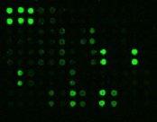
This paper focuses on the mechanism of epithelial-to-mesenchymal transition (EMT), which is an important process in tumor formation. The Human L-Series 507 Array yielded a short list of targets, including the TGFβ and WNT/β-catenin pathways, which are known to antagonize one another. By knowing where to look, they were able to focus their efforts on the right signal pathways, greatly improving their chances of working out the entire mechanism of this process, which netted them a paper in Cell.
Using a cytokine array keeps you from missing something important.
Vargas DL, Nascimbene C, Krishnan C, Zimmerman AW, Pardo CA. Neuroglial activation and neuroinflammation in the brain of patients with autism. Annals of Neurology. 2005; 57(1):67–81.
The authors of the paper found the first evidence of a neuroinflammatory process in the pathology of autism, even identifying several key factors tha t pointed to the mechanism of this process. This neuroinflammatory component of autism had been suspected for years, but no one had ever proven it. If the authors had decided to select a few ELISA kits or even a 20-plex bead panel to do this investigation instead of an array kit detecting 120 different cytokines, the authors may have missed identifying the key targets, and we might still be wondering if autism has a neuroinflammatory component.
Cell–cell signaling pathways are complex, and multiple factors may be working together, in concert.Therefore, screening for a limited number of secreted targets will often give you an incomplete picture.
Coppé J-P, Patil CK, Rodier F, Sun Y, Muñoz DP, Goldstein J, Nelson PS, Desprez P-Y, Campisi J. Senescence-associated secretory phenotypes reveal cell-nonautonomous functions of oncogenic RAS and the p53 tumor suppressor. PLoS Biology. 2008; 6(12).
This paper describes the first characterization of the senescence-associated secretory phenotype in stromal–tumor interactions. It uncovers the expression profile of permanently senescent tissues (epithelial and stromal) that secrete a wide range of factors that included pro-inflammatory factors, growth factors, cell-adhesion factors and proteases that restructure the extracellular matrix (ECM). In subsequent inquiries, these various components of this broad secretory profile were shown to work in concert to promote tumorigenesis. The Human Cytokine Array 1000 was instrumental in this discovery.
【01】 定量的抗体アレイ(Quantibody)と半定量的抗体アレイ(C、G、Lシリーズ)の違いは何ですか?
定量的アレイではアナライトの実際の濃度データ(例:pg/ml)を得ることができる一方、半定量的アレイでは2つ以上のサンプル間におけるアナライトの相対的変化についてデータを得られます。
【02】 膜ベースとガラススライドベースの抗体アレイの違いは何ですか?
違いはいくつかありますが、主な違いとしては検出方法です。ガラススライドベースの抗体アレイは、蛍光検出のため、互換性のある蛍光スキャナーが必要です。( 互換性のある/推奨される蛍光スキャナーのリストについてはこちらを参照ください。 )
一方、膜ベースの抗体アレイは、化学発光検出のため、ほとんどのウェスタンブロットイメージングシステムが適合します。
必要なサンプル量についても異なります。膜ベースの抗体アレイに対して、ガラススライドベースの抗体アレイの方が少ないサンプル容量で実施できます。(ガラススライドの場合:〜70-100 ul(希釈後) 膜ベースの場合:最低 1 ml(希釈後))
【03】 抗体アレイを行うにあたり、どのような機器が必要になりますか?
膜ベースの抗体アレイの場合、X線フィルム現像液、 CCDカメラ、またはゲルドキュメンテーションシステムなどの任意の化学発光イメージングシステムが必要です。(例えばLi-Cor Odyssey や Typhoon systemsなどの、近赤外蛍光のイメージングシステムがRBT社の膜ベースアレイでよく機能します。)
ガラススライドベースの抗体アレイの場合、遺伝子マイクロアレイ蛍光スキャナーが必要です。
互換性のある/推奨される蛍光スキャナーのリストについてはこちらを参照ください。
【04】 適用サンプルの種類は何になりますか?
ほとんどのアレイは、あらゆる生体液試料で使用できます。生体液試料とは、細胞培養培地、細胞ライセート、組織ライセート、およびすべての体液上清(血清、血漿、尿、脳脊髄液、気管支肺胞洗浄液:BAL、唾液、涙など)を含みます。
例外的に、ラベルベースの「L-シリーズ」では、血清/血漿以外の生体液試料やライセートにはご使用いただけません。
また、膜結合型受容体を検出するリン酸化抗体アレイは、細胞と組織ライセートサンプルでのみ検証されており、それらのサンプルでの使用を推奨します。
通常は、RBT社抗体アレイは水溶性・非変性タンパク質を含むセルフリー抽出液であれば実施できます。
下記よりそのほかのFAQもご覧いただけます。


 このページを印刷する
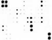


 中身を見る
中身を見る 中身を見る
中身を見る 中身を見る
中身を見る
 中身を見る
中身を見る