


 このページを印刷する
MITO-ID® Red / Green 検出キットは、生細胞、固定後の細胞の多重染色に最適なミトコンドリア染色キットです。
記事ID : 7428
ミトコンドリアの膜電位に関係なく、光安定且つ非毒性で、選択的に染色 MITO-ID® Red / Green ミトコンドリア染色キット
使用目的
MITO-ID® Red / Green 検出キットはエネルギー状態 (ミトコンドリア膜電位) に関係なく、ミトコンドリアを染色します。この色素は、従来の共焦点蛍光顕微鏡やハイコンテンツスクリーニング (HCS) プラットフォームなど、ほとんどの蛍光検出システムと互換性があり、蛍光タンパク質との蛍光共局在イメージング、ミトコンドリアの形態変化の評価、質量の推定に役立ちます。
特長
- 生細胞、界面活性剤で透過した細胞、アルデヒド固定した細胞の何れも染色可能
- 多重染色に最適
- 光退色に高い耐性あり
構成内容
- MITO-ID® Red 検出試薬(Ex/Em=558nm/690nm)もしくは MITO-ID® Green 検出試薬(Ex/Em=460nm/560nm)
- Hoechst 33342 核染色試薬
- 10X アッセイバッファー
アプリケーション例

図1 HeLa細胞のイメージング像
細胞を MITO-ID® Red ミトコンドリア染色キット(品番51007-500)で15分間染色した。核を Hoechst 33342 で対比染色した。
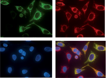
図2 MITO-ID® Red 検出キットによるHeLa-TurboGreen-ミトコンドリア細胞株 (GFP-チトクロームcオキシダーゼを発現) の染色画像
左上:未処理、右上:MITO-ID® Red 検出試薬処理、左下:Hoechst 33342 処理、右下:合成画像。MITO-ID® Red はEGFP-チトクロームcオキシダーゼシグナル (黄色またはオレンジのシグナル) と共局在し、ミトコンドリアの選択性を示す。GFPタグ付きタンパク質を発現しなくなった細胞内のミトコンドリア、または量が減少したものは、合成画像で赤く示されている。
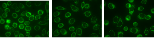
図3 ミトコンドリアの膜電位に影響を受けにくい MITO-ID® Green色素による染色画像
左:MITO-ID® Green色素による生細胞のミトコンドリアの選択的染色、中央:脱文局在である CCCP (シアン化カルボニル3-クロロフェニルヒドラゾン) 処理、右:イオンチャネル摂動薬である valinomycin 処理
テックノート
MITO-ID® Red ミトコンドリア染色キット
| 品名 | メーカー | 品番 | 包装 | 希望販売価格 | 在庫 |
|---|---|---|---|---|---|
MITO-ID(R) Red detection kit (GFP CERTIFIED(R)) |
ENZ | ENZ-51007-500 | 1 KIT [500 assays] |
¥74,000 | なし |
| MITO-ID(R) Red detection kit (GFP CERTIFIED(R)) |
ENZ | ENZ-51007-0100 | 100 TEST |
¥26,000 | あり |
MITO-ID® Green ミトコンドリア染色キット
| 品名 | メーカー | 品番 | 包装 | 希望販売価格 | 在庫 |
|---|---|---|---|---|---|
| MITO-ID(R) Green detection kit |
ENZ | ENZ-51022-K500 | 1 KIT [500 assays] |
¥74,000 | なし |
| MITO-ID(R) Green detection kit |
ENZ | ENZ-51022-0100 | 100 TEST |
¥26,000 | なし |
製品使用文献
MITO-ID® Red ミトコンドリア染色キット(品番: ENZ-51007-500 / ENZ-51007-0100)
- Effects of two fullerene derivatives on monocytes and macrophages: S. Pacor, et al.; Biomed. Res. Int. (2015), Application(s): Confocal microscopy on monocytes and macrophages, Abstract;
 Full Text
 - Induction of androgen formation in the male by a TAT-VDAC1 fusion peptide blocking 14-3-3? protein adaptor and mitochondrial VDAC1 interactions: Y. Aghazadeh, et al.; Mol. Ther. 22, 1779 (2014), Abstract;
- Protein modifications regulate the role of 14-3-3γ adaptor protein in cAMP-induced steroidogenesis in MA-10 Leydig cells: Y. Aghazadeh, et al.; J. Biol. Chem. 289, 26542 (2014), Abstract;
- Mitochondrial fission induced by platelet-derived growth factor regulates vascular smooth muscle cell bioenergetics and cell proliferation: J.K. Salabei, et al.; Redox Biol. 1, 542 (2013), Application(s): Detection by confocal microscopy, Abstract; Full Text
- Utilization of fluorescent probes for the quantification and identification of subcellular proteomes and biological processes regulated by lipid peroxidation products: T.D. Cummins, et al.; Free Radic. Biol. Med. 59, 56 (2013), Application(s): Detection by confocal microscopy, Abstract; Full Text
- Ectopic ATP synthase blockade suppresses lung adenocarcinoma growth by activating the unfolded protein response: H.Y. Chang, et al.; Cancer Res. 72, 4696 (2012), Abstract; Full Text
- Hormone-induced 14-3-3γ adaptor protein regulates steroidogenic acute regulatory protein activity and steroid biosynthesis in MA-10 Leydig cells: Y. Aghazadeh, et al.; J. Biol. Chem. 287, 15380 (2012), Application(s): Detection of mitochondria in MA-10 mouse Leydig tumor cells using confocal microscopy, Abstract; Full Text
- Intracellular Energetic Units regulate metabolism in cardiac cells: V. Saks, et al.; J. Mol. Cell. Cardiol. 52, 419 (2012), Application(s): Detection of mitochondria in cardiomyocytes using confocal microscopy, Abstract;
- Opa3, a novel regulator of mitochondrial function, controls thermogenesis and abdominal fat mass in a mouse model for Costeff syndrome: T. Wells, et al.; Hum. Mol. Genet. 18, 4836 (2012), Application(s): Visualization of mitochondria in paraffin embedded sections of mouse brown adipose tissue with Mito-ID® Red detection kit., Abstract; Full Text
- Cytometric assessment of mitochondria using fluorescent probes: C. Cottet-Rousselle, et al.; Cytometry A. Jun 79(6), 405 (2011), Abstract;
- Mitochondria-cytoskeleton interaction: distribution of β-tubulins in cardiomyocytes and HL-1 cells: R. Guzun, et al.; Biochim. Biophys. Acta 1807, 458 (2011), Application(s): Detection of mitochondria in cardiomyocytes using fluorescence microscopy, Abstract;
- The targeting of plasmalemmal ceramide to mitochondria during apoptosis: E.B. Babiychuk, et al.; PLoS One 6, e23706 (2011), Application(s): Detection of mitochondria in T cell and monocyte cell lines using fluorescence microscopy, Abstract; Full Text
MITO-ID® Green ミトコンドリア染色キット(品番: ENZ-51022-K500 / ENZ-51022-0100)
- A new and reliable method for live imaging and quantification of reactive oxygen species in Botrytis cinerea: technological advancement: R. Marschall, et al.; Fungal Genet. Biol. 71, 68 (2014), Abstract;
参考文献
- Photoconversion of Lysotracker Red to a green fluorescent molecule: E.C. Freundt, et al.; Cell Res. 17, 956 (2007), Abstract;
- Systematic colocalization errors between acridine orange and EGFP in astrocyte vesicular organelles: F. Nadrigny, et al.; Biophys. J. 93, 969 (2007), Abstract;
- Chloromethyl-X-rosamine (MitoTracker Red) photosensitises mitochondria and induces apoptosis in intact human cells: T. Minamikawa, et al.; J. Cell. Science 112, 2419 (1999), Abstract;
- Chloromethyltetramethylrosamine (Mitotracker Orange) induces the mitochondrial permeability transition and inhibits respiratory complex I. Implications for the mechanism of cytochrome c release: L. Scorrano, et al; J. Biol. Chem. 274, 24657 (1999), Abstract;
関連商品
- ミトコンドリア染色試薬
生細胞/固定細胞染色用の各種色素を販売
(JC-1/MitoView 633/Rhodamine 123/TMRE/TMRM など) - Cell Navigator® ミトコンドリア染色キット
3色の蛍光色素から選択可能!生細胞内のミトコンドリアを蛍光標識
商品は「研究用試薬」です。人や動物の医療用・臨床診断用・食品用としては使用しないように、十分ご注意ください。
※ 表示価格について

- 「MITO-ID® Red / Green ミトコンドリア染色キット」は、下記のカテゴリーに属しています。