


 このページを印刷する
NUCLEAR-ID® Red DNA 染色色素は、蛍光検出技術で用いられるように設定された、核を識別するための細胞透過性色素です。蛍光退色に耐性を持ち、生細胞の核染色に最適です。また、フローサイトメトリーによる細胞周期進行の誘導や阻害研究に便利です。生細胞での潜在的な用途として、細胞内DNA量の決定、細胞周期進行の決定、成長パターンのバリエーション決定、アポトーシス観察、評価腫瘍細胞の動向、抑制遺伝子メカニズムなどがあります。
特長
- DNAを特異的に染色する遠赤色蛍光色素
- 高純度で高い安定性
- 生細胞/透過処理した細胞/固定した細胞の解析が可能
- 光退色なし
- RNase 処理は不要
- GFP および FITC との多重染色が可能
- UVレーザーによる励起は不要
- 様々な細胞密度で検証済み
- 短時間で簡単に染色
その他
波長 (Ex/Em nm): 566/650
使用例
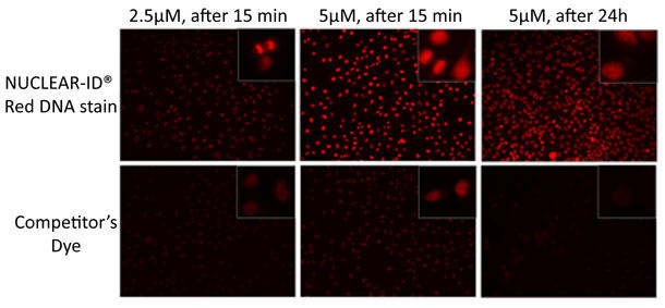
NUCLEAR-ID® Red DNA 色素は他社競合品よりも低濃度で dsDNA を可視化。
HeLa細胞は 〜 60% コンフルエントになるまで培養した。それから本試薬もしくは競合品で染色(濃度2.5µM、5.0µM、37℃)し、やさしく洗浄した。写真は、15分後、24時間後に撮影。結果は、2.5µM NUCLEAR-ID® Red DNA 色素では dsDNA の可視化が可能で、他社競合品では 5.0µM を要した。24時間後では、他社競合品の強度や細胞成長は、終濃度 5.0µM で著しく減少した。同時間、濃度での NUCLEAR-ID® Red DNA 色素では、毒性が低く、生細胞研究には、より低濃度で行えることが分かった。
NUCLEAR-ID® Red DNA 染色色素
| 品名 | メーカー | 品番 | 包装 | 希望販売価格 |
|---|---|---|---|---|
NUCLEAR-ID(R) Red DNA Stain |
ENZ | ENZ-52406 | 200 UL |
¥89,000 |
【関連商品】
- Nucleolar-ID® Green 検出キット
- 生きた細胞の核小体を染色、動態変化を観察可能 - TOTAL-NUCLEAR-ID® green/red nucleolar/nuclear 検出キット
- 生細胞の核小体と核を同時染色 - NUCLEAR-ID® Blue DNA 染色色素 (GFP-CERTIFIED®)
- 幅広いアプリケーションに使用できる細胞透過性DNA染色色素
製品使用文献
- Cellular IAP proteins and LUBAC differentially regulate necrosome-associated RIP1 ubiquitination: M.C. de Almagro, et al.; Cell Death Dis. 6, e1800 (2015), Abstract;
 Full Text
 - Plasminogen activator inhibitor-1 regulates tumor initiating cell properties in head and neck cancers: Y.C. Lee, et al.; Head Neck (2015), Abstract;
- Pervasive axonal transport deficits in multiple sclerosis models: C.D. Sorbara, et al.; Neuron 84, 1183 (2014), Abstract;
- Zscan4 is regulated by PI3-kinase and DNA-damaging agents and directly interacts with the transcriptional repressors LSD1 and CtBP2 in mouse embryonic stem cells: M.P. Storm, et al.; PLoS One 9, e89821 (2014), Application(s): Flow cytometry measurements on mouse pluripotent cells, Abstract; Full Text
- Epigenetic regulation of planarian stem cells by the SET1/MLL family of histone methyltransferases: A. Hubert, et al.; Epigenetics 8, 79 (2013), Application(s): Cell cycle analysis by flow cytometry, Abstract; Full Text
- Wntless is required for peripheral lung differentiation and pulmonary vascular development: B. Cornett, et al.; Dev. Biol. 379, 38 (2013), Application(s): Detection of nuclei in lung cells by flow cytometry, Abstract;
- Ionizing radiation induces mitochondrial reactive oxygen species production accompanied by upregulation of mitochondrial electron transport chain function and mitochondrial content under control of the cell cycle checkpoint: T. Yamamori, et al.; Free Radic. Biol. Med. 53, 260 (2012), Application(s): Cell cycle analysis by flow cytometry, Abstract;
- Neurokinin 1 receptor mediates membrane blebbing and sheer stress-induced microparticle Formation in HEK293 Cells: P. Chen, et al.; PLoS One 7, e45322 (2012), Application(s): DNA nuclei staining by flow cytometry, Abstract; Full Text
- Synthesis, cytotoxicity and cellular uptake studies of N3 functionalized Re(CO)3 thymidine complexes: M.D. Bartholomä, et al.; Dalton Trans. 40, 6216 (2011), Application(s): Nuclear DNA stain of human lung adenocarcinoma cells, Abstract;
- A cell-permeant dye for cell cycle analysis by flow and laser-scanning microplate cytometry: Y.J. Xiang, et al.; Nat. Methods 6, an2 (2009), Application(s): Cell cycle analysis by flow cytometry, Abstract;
商品は「研究用試薬」です。人や動物の医療用・臨床診断用・食品用としては使用しないように、十分ご注意ください。
※ 表示価格について

- 「NUCLEAR-ID® Red DNA 染色色素」は、下記のカテゴリーに属しています。


 中身を見る
中身を見る 中身を見る
中身を見る 中身を見る
中身を見る
 中身を見る
中身を見る