For Research Use Only No Crystallization Necessary
Providing Protein Molecular Structural Analysis Service
Background
Knowing the protein structure became crucial for life sciences these days. The new, unique technology called Protein Tomography enables effective drug discovery by revealing critical information at early stages of drug development. It allows visualization of protein tertiary structures at the individual, single molecule level in short time with high accuracy. Proteins that with conventional methods were unable to visualize, are now easily reconstructed with our service, Protein Tomography.
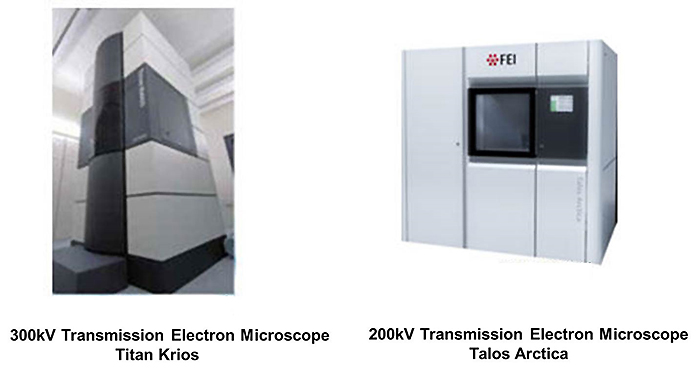
The samples, first vitrified, are set in a sophisticated Cryo Electron Microscope called Titan Krios, where cross section images are taken. Next, the tomograms are aligned, creating three dimensional structure. The structure then is refined with specifically designed program called COMET (Constrained Maximum Entropy Tomography), which innovative algorithms enhance resolution, producing noise free reconstructions of target proteins. Biopharmaceutical companies related to drug discovery and development, as well as academic and research institutions are to benefit most from this innovative technology. Located in the southern island of Japan, Okinawa Protein Tomography provides this unique service worldwide.
Service features
- Unique combination of Cryo Electron Microscopy and specifically designed reconstruction program COMET allowing three dimensional macromolecular analysis (no crystallization necessary)
- Three dimensional macromolecular reconstruction with a higher success rate when compared with the traditional methods.
- Ability to grasp the dynamics of single proteins by analyzing structures of separate, individual molecules as opposed to the conventional averaging technologies
- Visualization close to in vivo state, through vitrification, without staining and/or substitution
- Minimum amount of the sample starts from 1µl (e.g. 3 vials with 10µl if concentration is 1mg/ml)
- Applicable to proteins and protein complexes impossible to visualize with other methods
What Is Tomography?
- A method applying principle of cross section imaging in transmission electron microscopy
- Creating 3D reconstruction by aligning the cross section images
Advantages of Protein Tomography
- Possibility of obtaining protein structure information at the domain level even in case of the samples, which failed imaging with other methods, e.g. crystallography
Clarity of binding area and molecular association - Possibility of building structural classes and calculating the ratio of identified multimers
Possibility of analyzing diverse molecular associations and structural states of target proteins
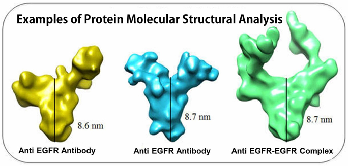
Increasing the number of assayed molecules allows revealing the conformational distribution and apprehending the structural characteristics of each association and state - Possibility of analyzing structural differences of separate, individual molecules, since the technology is not using any of the averaging methods
By comparing different yet biologically identical molecules, it is possible to reveal the reason behind low activity or reactivity differences - Possibility of using same protein solution as for crystallography (unused surplus)
Comparison of results generated by different methods (e.g. crystallography and Protein Tomography) became possible along with imaging the protein solutions inapplicable for crystallization - Possibility of comparing the results obtained from Protein Tomography with other methods
Analyzing individual molecules (HS-AFM imaging etc.) or structural analysis of proteins in solution (SAXS etc.) offer new insight into the field of molecular imaging
It is also possible to verify the structural distribution combining Native PAGE, MALDI-TOF/MS and/or gel filtration chromatography and others.
Our process
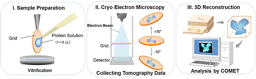
- Sample Preparation
Samples are vitrified to catch the molecules in the closest to in vivo state possible, without staining and substitution. Even small amount of the sample can be analyzed. Minimum amount of the sample starts from 1µl (e.g. 3 vials with 10µl if concentration is 1mg/ml) - Collecting Data from Cryo Electron Microscope
High performance direct detector and reduction of electron beam prevents the molecules from degeneration. - 3D Reconstruction
Analysis by COMET (Constrained maximum entropy tomography). Enhancing the tomograms by improving the ratio of signal/noise
This product is "Research reagent". It is prohibited to use against humans or animals in medical care, clinical diagnoses, or as food.