ページの本文へ移動

研究用

生細胞を安定して標識し、細胞追跡実験に最適 CYTO-ID® 長期細胞トレーサーキット

このエントリーをはてなブックマークに追加

細胞の脂質二重膜に非共有結合する独自の蛍光色素を用いた細胞膜染色キットです。
長期的な細胞の生存率、毒性、接着、遊走、融合等を調べる研究に最適です。

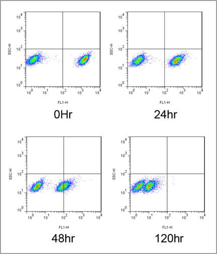
本キットは、独自の非共有結合細胞標識技術を使用して、疎水性脂肪族鎖を含む蛍光色素を細胞膜の脂質二重層に安定的に組み込みます。蛍光色素は、細胞にロード後最大96時間保持され、有糸分裂時には娘細胞に引き継がれます。色素は細胞内のタンパク質を共有結合的に修飾しないため、チオール反応性クロロメチルベースまたはアミン反応性スクシンイミジルエステルベースの蛍光色素をベースとした分子プローブを使用するよりも、通常の生理学的反応がよりよく保存されます。

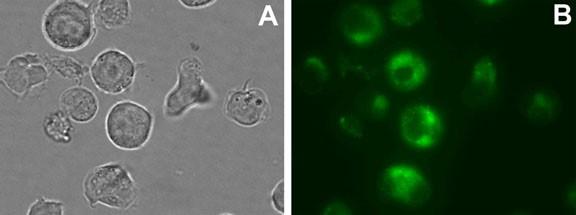
標識された細胞は、細胞の種類によって、均一に明るいものから小さな顆粒状(punctate)に染色されるものまでさまざまです。この違いは、細胞標識後に生じる膜の内在化の程度に関連すると考えられています。 CYTO-ID®Greenトレーサー色素の蛍光は、通常の生理学的範囲内のpHに依存することなく、細胞ごとの蛍光強度は色素分布のパターンの影響を受けません。標識した細胞は、蛍光顕微鏡または共焦点蛍光顕微鏡で観察でき、さらに、フローサイトメトリーにより色素標識および非標識細胞集団を分析することも可能です。

また、色素ロード後96時間のインキュベーション後にも、隣接細胞への蛍光の移動は観察されませんでした。これは、生理的温度で数時間生存細胞内にのみ保持されるカルセインAMおよびBCECF AMとはまったく対照的です。

特長

  • 蛍光顕微鏡やフローサイトメーターで観察・検出ができます。
  • 細胞毒性はありません。
  • 染色96時間後でも蛍光が安定していて、有糸分裂後の娘細胞においても蛍光観察が可能です。
  • 隣接した細胞への蛍光の転移はありません。
  • 細胞内の生理学的なpH変化に影響を受けません。
  • 染色試薬はタンパク質に共有結合しないため、チオール反応性クロロメチル基やアミン反応性スクシンイミジルエステル基を有する蛍光色素を用いた分子プローブに比べ、正常な生理学的条件下で観察できます。
  • Texas Red®用のフィルターが使用できます。
  • 二重標識が可能です。

構成内容

  • Cyto-ID® Red Tracer Dye
  • 標識バッファー(×4)
  • HBSS(×10)

使用例

long-term-cell-tracer-kits-enz_01.jpg

図1. Jurkat細胞の染色画像
(A) 明視野  (B) CYTO-ID® Green Tracer 色素による染色画像
標準的なFITCフィルターを使用して、膜結合蛍光を観察した。

long-term-cell-tracer-kits-enz_02.jpg

図2. Jurkat細胞混合集団の経時的蛍光のフローサイトメトリー解析
CYTO-ID®Green Tracer色素で染色したJurkat細胞を、染色していないJurkat細胞集団と混合し、120時間培養した。

CYTO-ID® 長期細胞トレーサーキット

品名 メーカー 品番 包装 希望販売価格
CYTO-ID(R) Green long-term cell tracer kit詳細データ ENZ ENZ-51036-K025 1 KIT
¥56,000
CYTO-ID(R) Red long-term cell tracer kit詳細データ ENZ ENZ-51037-K025 1 KIT
[25 assays]
¥53,000

【関連商品】

製品使用文献

  1. Proangiogenic effects of tumor cells on endothelial progenitor cells vary with tumor type in an in vitro and in vivo rat model. R. An, et al. FASEB J. 32: 5587 (2018), Application(s): Identification of TR-BME2 cells in tube-formation co-culture assay with CC521 colon adenocarcinoma cells.
  2. A liquid biopsy-based method for the detection and quantification of circulating tumor cells in surgical osteosarcoma patients. H. Zhang, et al. Int. J. Oncol. 50: 1075 (2017), Application(s): Use with HOS cells (osteosarcoma). Microscopy
  3. Characterization and Cellular Internalization of Spherical Cellulose Nanocrystals (CNC) into Normal and Cancerous Fibroblasts. N.A.H. Shazali, et al. Materials (Basel). 12: 3251 (2019), Application(s): C6 and NIH3T3 cell tracing
  4. Time resolved 3D live-cell imaging on implants. A. Ingendoh-Tsakmakidis, et al. PLoS One. 13: e0205411 (2018)
  5. Nerve guidance conduit with a hybrid structure of a PLGA microfibrousbundle wrapped in a micro/nanostructured membrane: S.W. Peng, et al. Int. J. Nanomedicine. 12: 421 (2017), Application(s):Tracking in vitro cells migration (immortalized neuron progenitor KT 98)
  6. Thioreductase-containing epitopes inhibit the development of type 1 diabetes in the NOD mouse model. E. Malek Abrahimians, et al. Front. Immunol. 7, 67 (2016), Application(s): Flow cytometry analysis using isolated mouse spleen B cells
  7. Syngeneic murine ovarian cancer model reveals that ascites enriches for ovarian cancer stem-like cells expressing membrane GRP78. L. Mo, et al. Mol. Cancer Ther. 14: 747 (2015)
  8. An in vitro blood-brain barrier model combining shear stress and endothelial cell/astrocyte co-culture. Y. Takeshita, et al. J. Neurosci. Methods. 232, 165 (2014), Application(s): Confocal microscopy using adult human astrocyte cells
  9. Vascular endothelial growth factor-C modulates proliferation and chemoresistance in acute myeloid leukemic cells through an endothelin-1-dependent induction of cyclooxygenase-2. K.T. Hua, et al. Biochim. Biophys. Acta. 1843: 387 (2014)
  10. Expanding the Ig superfamily code for laminar specificity in retina: expression and role of contactins. M. Yamagata, et al. J. Neurosci. 32: 14402 (2012)

商品は「研究用試薬」です。人や動物の医療用・臨床診断用・食品用としては使用しないように、十分ご注意ください。

お問い合わせ


メーカー・代理店一覧

サポート情報

SNSアカウント

オウンドメディア

※当社のWEBサイトはユーザーの利便性を最適にし、それを保証するためにクッキーを使用しています。
 このWEBサイトの利用を継続することで、クッキーの使用に同意することになります。

© COSMO BIO