■ CD8a モノクローナル抗体(品番:65144-1-Ig)
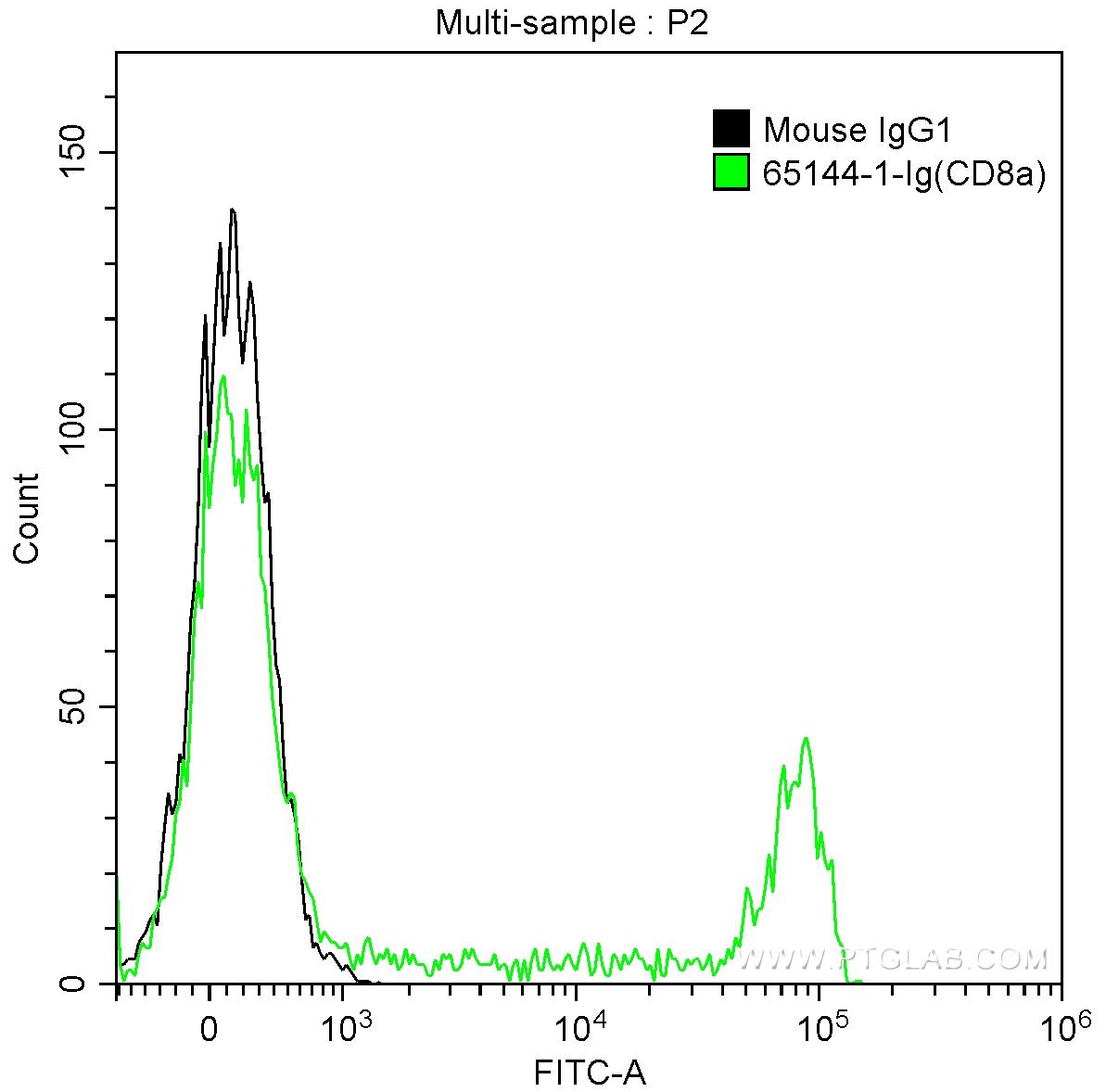
図1. CD8a抗体とアイソタイプコントロール抗体を用いた細胞表面マーカー解析
1X10^6 human peripheral blood lymphocytes were surface stained with 0.5 ug Anti-Human CD8a (65144-1-Ig, Clone:RPA-T8) and CoraLite®488-Conjugated AffiniPure Goat Anti-Mouse IgG(H+L) at dilution 1:1000 (green), or stained with 0.5 ug isotype control antibody and CoraLite®488-Conjugated AffiniPure Goat Anti-Mouse IgG(H+L) at dilution 1:1000 (black). Samples were not fixed.
■ CD8a モノクローナル抗体(品番:FITC-65144)
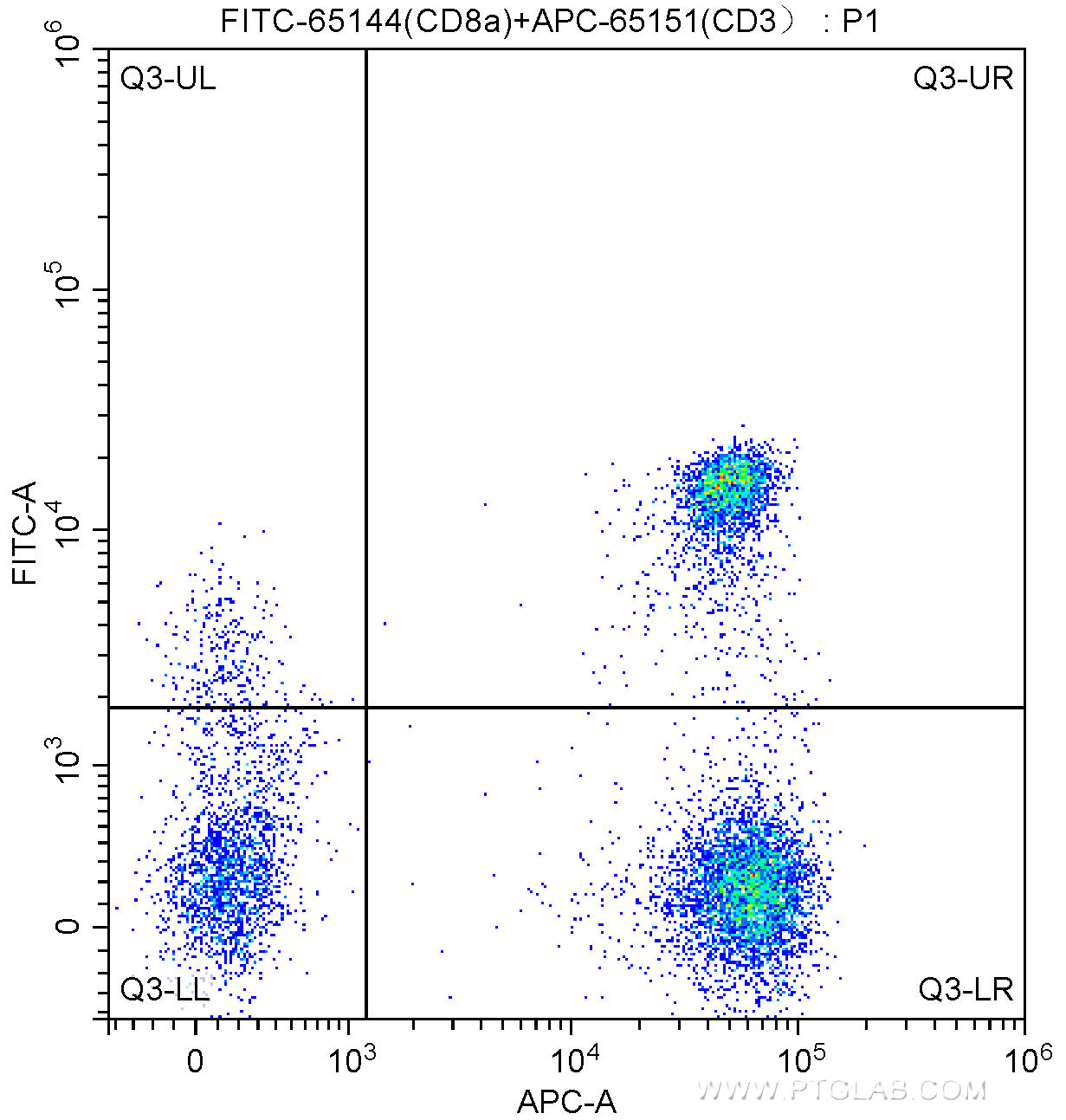
図1. CD8a抗体とCD3抗体を用いた細胞表面マーカー解析
100 ul human peripheral blood were surface stained with APC Anti-Human CD3 (APC-65151, Clone: UCHT1) and 5.00 ul FITC Anti-Human CD8a (FITC-65144, Clone: RPA-T8). Lymphocytes were gated for analysis. Cells were not fixed.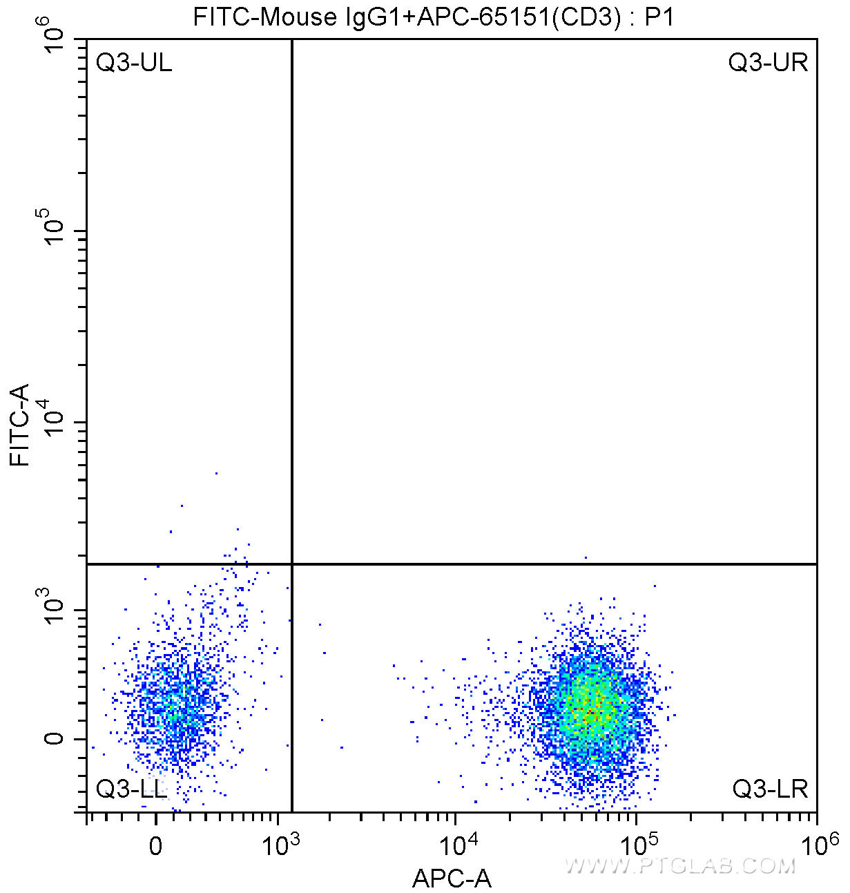
図2. アイソタイプコントロール抗体とCD3抗体を用いた細胞表面マーカー解析
100 ul human peripheral blood were surface stained with APC Anti-Human CD3 (APC-65151, Clone: UCHT1) and FITC Mouse IgG1 isotype control antibody. Lymphocytes were gated for analysis. Cells were not fixed.
■ CD8a モノクローナル抗体(品番:PE-65144)
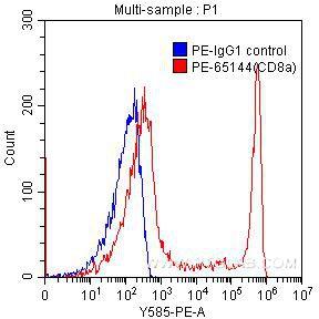
図1. CD8a抗体とアイソタイプコントロール抗体を用いた細胞表面マーカー解析
1X10^6 human peripheral blood lymphocytes were surface stained with 0.125 ug PE-anti-human CD8a (PE-65144, clone RPA-T8) (red) or 0.125 ug PE-mouse IgG1 isotype control (blue). Samples were not fixed.
■ CD8a モノクローナル抗体(品番:APC-65144)
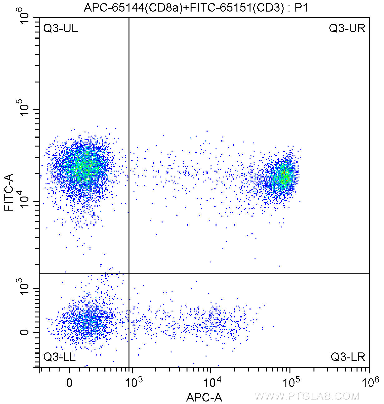
図1. CD8a抗体とCD3抗体を用いた細胞表面マーカー解析
100 ul human peripheral blood were surface stained with FITC Anti-Human CD3 (FITC-65151, Clone: UCHT1) and 5.00 ul APC Anti-Human CD8a (APC-65144, Clone: RPA-T8). Lymphocytes were gated for analysis. Cells were not fixed.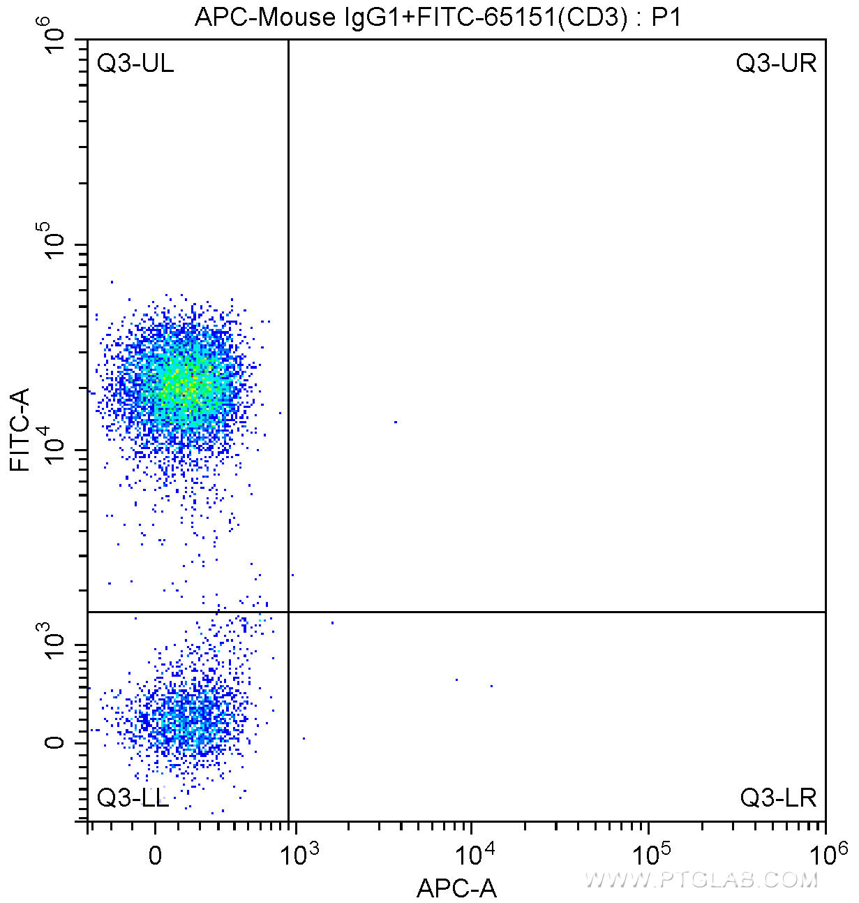
図2. アイソタイプコントロール抗体とCD3抗体を用いた細胞表面マーカー解析
100 ul human peripheral blood were surface stained with FITC Anti-Human CD3 (FITC-65151, Clone: UCHT1) and APC Mouse IgG1 isotype control antibody. Lymphocytes were gated for analysis. Cells were not fixed.
■ CD8a モノクローナル抗体(品番:CL488-65144)
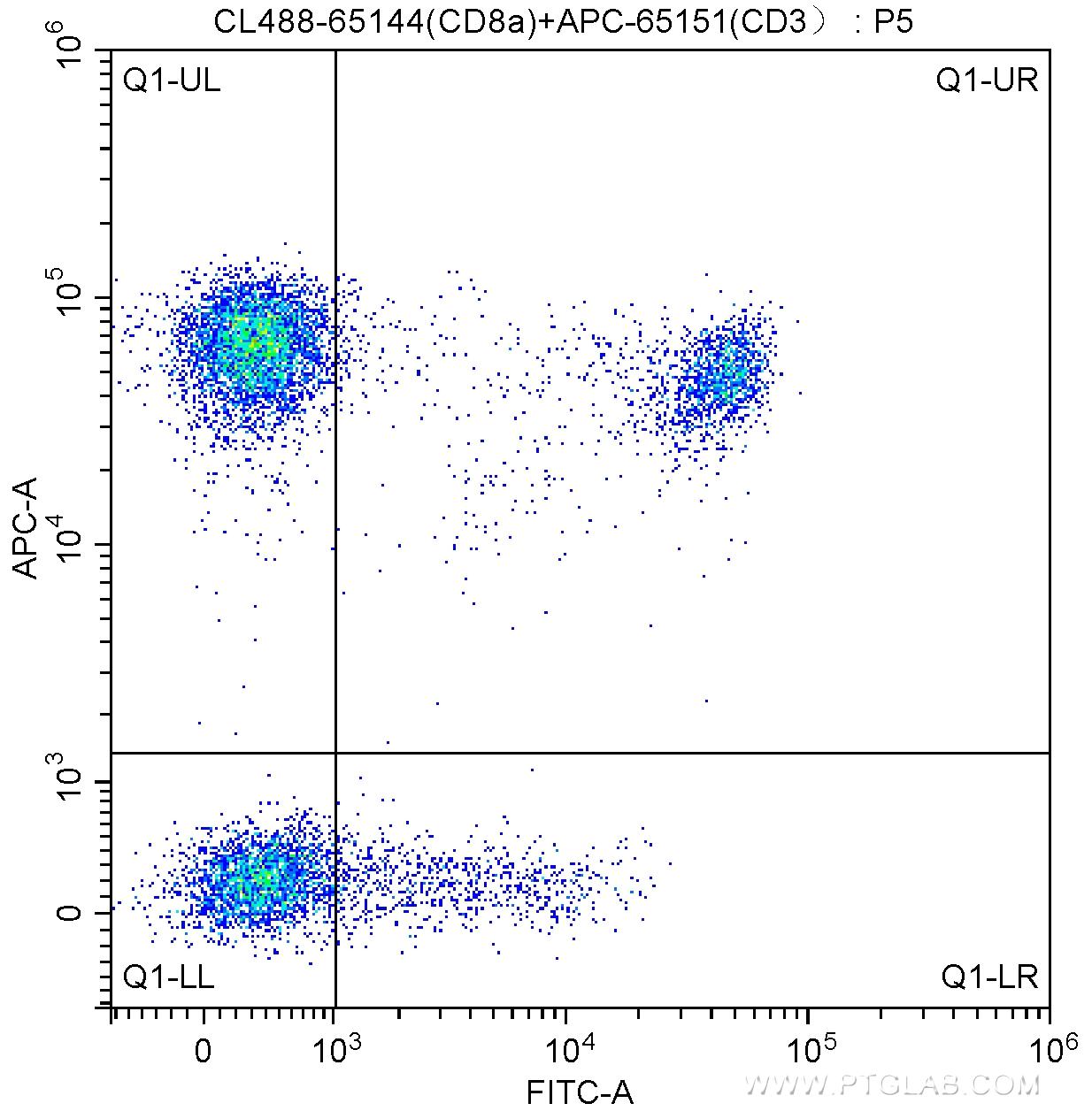
図1. CD8a抗体とCD3抗体を用いた細胞表面マーカー解析
100 ul human peripheral blood were surface stained with APC Anti-Human CD3 (APC-65151, Clone: UCHT1) and 5.00 ul CoraLite®488-conjugated Anti-Human CD8a (CL488-65144, Clone: RPA-T8). Lymphocytes were gated for analysis. Cells were not fixed.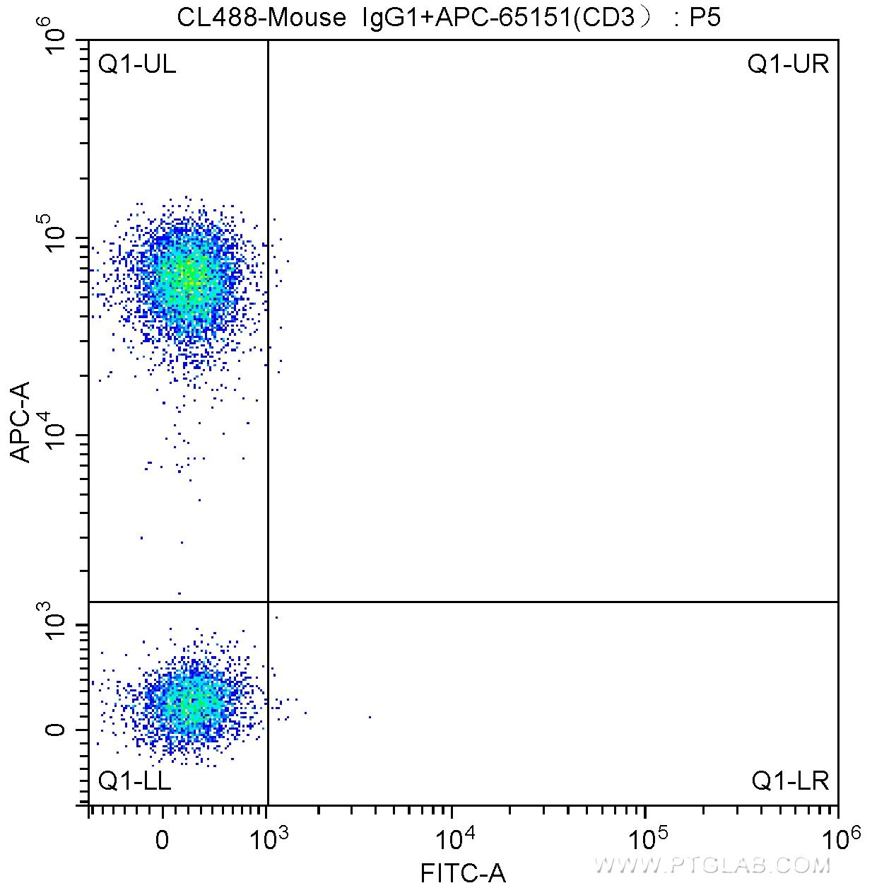
図2. アイソタイプコントロール抗体とCD3抗体を用いた細胞表面マーカー解析
100 ul human peripheral blood were surface stained with APC Anti-Human CD3 (APC-65151, Clone: UCHT1) and CoraLite®488-conjugated Mouse IgG1 isotype control antibody. Lymphocytes were gated for analysis. Cells were not fixed.
■ CD8a モノクローナル抗体(品番:CL647-65144)
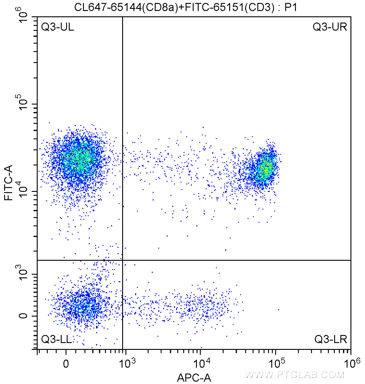
図1. CD8a抗体とCD3抗体を用いた細胞表面マーカー解析
100 ul human peripheral blood were surface stained with FITC Anti-Human CD3 (FITC-65151, Clone: UCHT1) and 5.00 ul CoraLite®647-conjugated Anti-Human CD8a (CL647-65144, Clone: RPA-T8). Lymphocytes were gated for analysis. Cells were not fixed.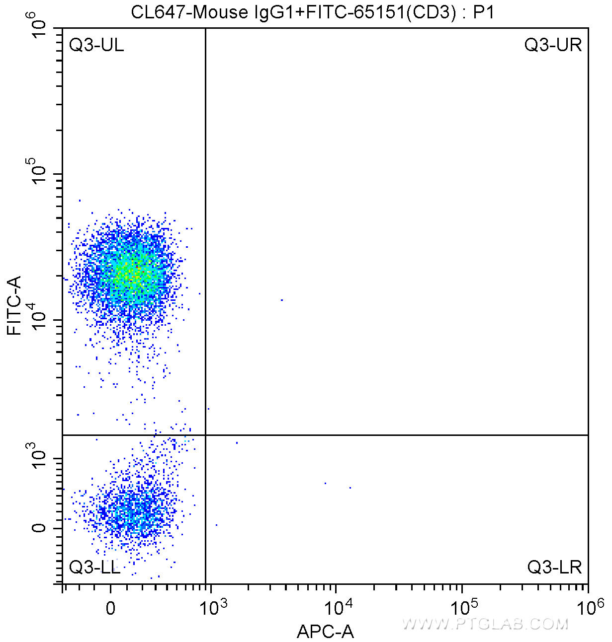
図2. アイソタイプコントロール抗体とCD3抗体を用いた細胞表面マーカー解析
100 ul human peripheral blood were surface stained with FITC Anti-Human CD3 (FITC-65151, Clone: UCHT1) and CoraLite®647-conjugated Mouse IgG1 isotype control antibody. Lymphocytes were gated for analysis. Cells were not fixed.
![]() または5点購入30%オフ専用申込書
または5点購入30%オフ専用申込書 ![]()


 このページを印刷する

