ページの本文へ移動

記事ID : 41836
研究用

高い浸透性!免疫細胞染色に最適、Nano-Label シリーズ Spot-Label®(蛍光標識Spotタグ抗体)

このエントリーをはてなブックマークに追加

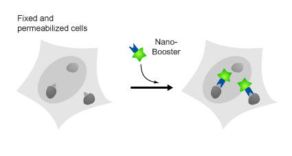
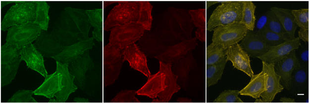
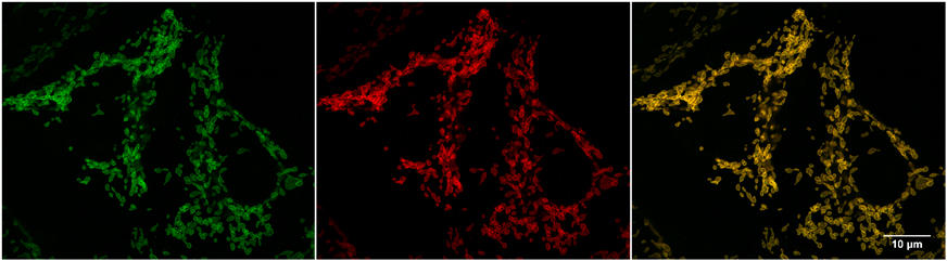
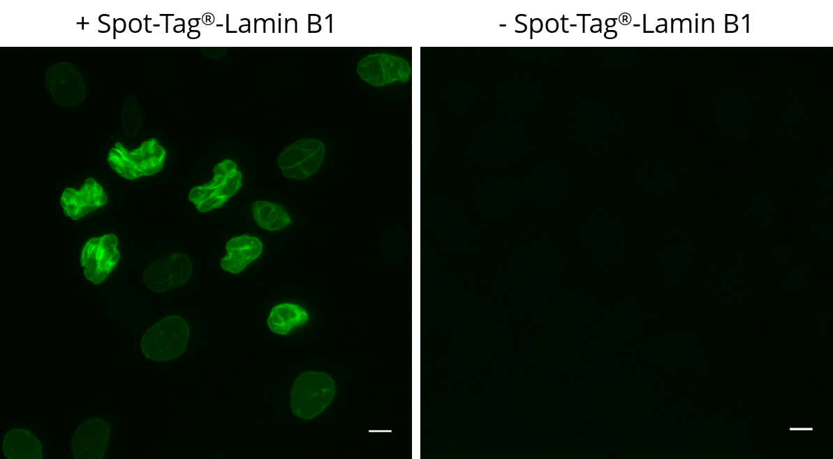
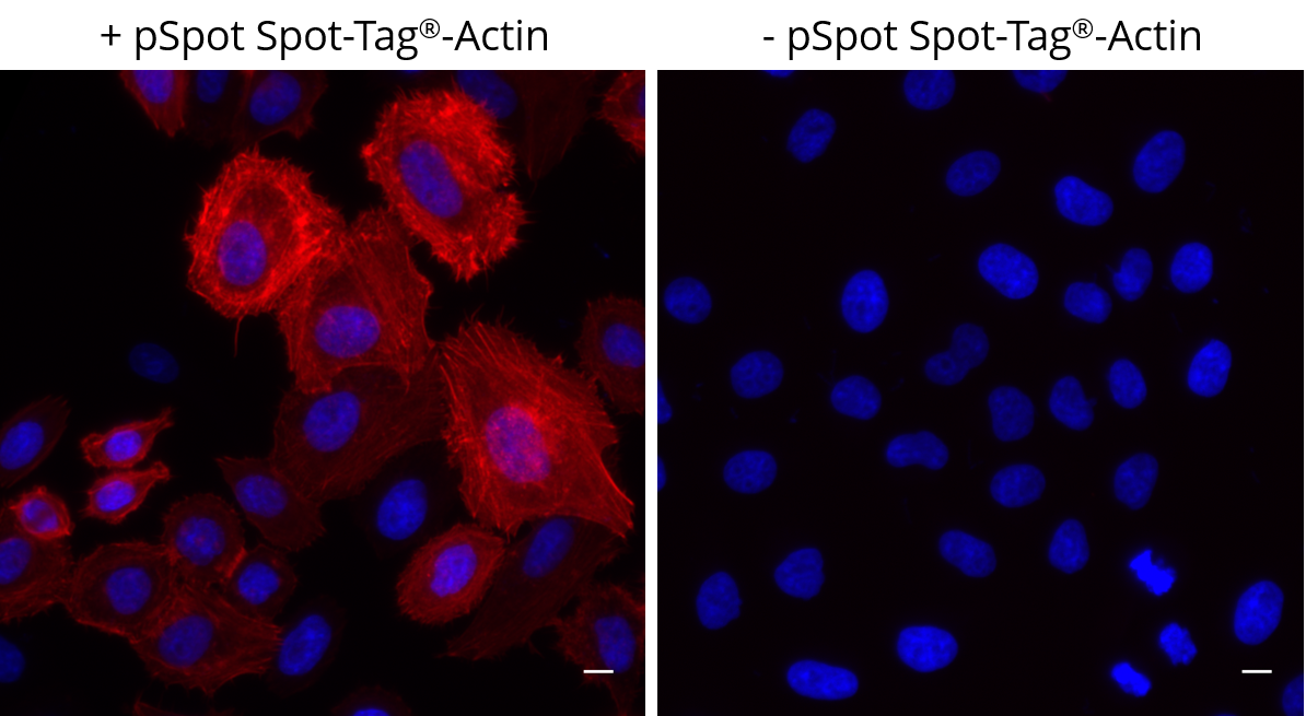
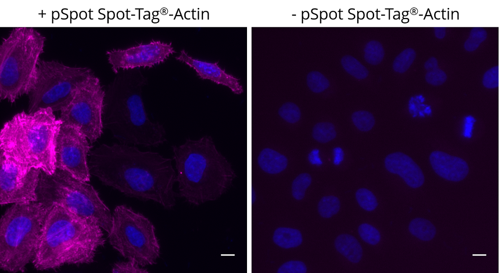
Spot-Label®(Spotラベル®)は、Spotタンパク質の免疫蛍光染色(IF:Immunofluorescence)実験に最適な蛍光色素標識済みのアルパカ由来Spotタグ抗体です。

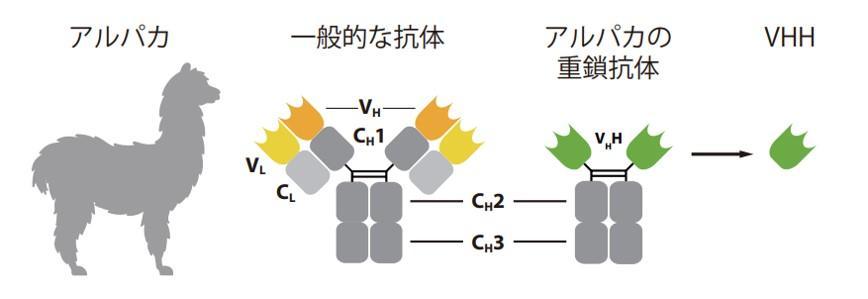
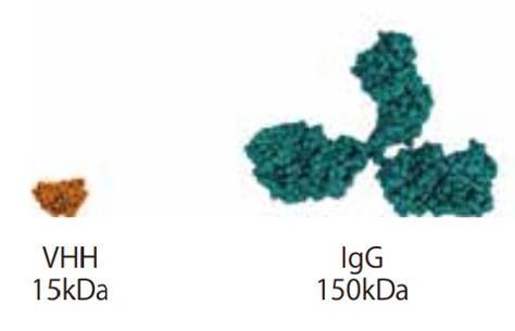
蛍光色素には、アルパカ(Alpaca)を宿主動物(免疫動物)として作製された「抗Spot組換えモノクローナルVHH抗体」が結合しています。VHH抗体は、Nanobody® としても知られるラクダ科動物の「重鎖抗体(Heavy chain-only antibody)」由来の抗原結合ドメインであり、高い特異性と親和性を示します。VHH抗体は、一般的の抗体よりもサイズが小さく浸透性が高いため、細胞染色に最適です。

高浸透性&高親和性アルパカ組換えVHH免疫蛍光染色(IF)に最適

背景

■ VHH抗体とは?

VHH抗体は、Nanobody® としても知られるラクダ科動物の「重鎖抗体(Heavy chain-only antibody)」由来の抗原結合ドメインです。一般的な抗体と比べて、10分の1程度とサイズが小さく、高い特異性と親和性を示すことで知られます。

図. ラクダ科動物のユニークな重鎖抗体・VHH
図. 従来の1/10ほどの分子量
図. 従来の1/10ほどの分子量
 

■ NanoBooster®/NanoLabel®とは?

Nano-Booster(ナノブースター)およびNano-Label(ナノラベル)は、蛍光タンパク質や細胞内タンパク質を標識する蛍光プローブです。蛍光色素(Alexa Fluor®またはAtto色素)に共有結合したVHH抗体で構成されます。抗体サイズが小さいため、高浸透性かつ高解像なイメージングに寄与し、免疫蛍光染色(IF)を中心としたアプリケーションに利用できます。

  • IF(免疫蛍光染色)/ ICC(免疫細胞化学)
  • IHC(免疫組織化学)
  • 超解像顕微鏡観察(SRM:Super-resolution microscopy)
図. Nano-Boosterの概要。GFP/RFP融合タンパク質の蛍光シグナルは、細胞固定後に減少します。GFP/RFP-Booster®は、蛍光シグナルの増幅に利用できます
図. Nano-Boosterの概要。
GFP/RFP融合タンパク質の蛍光シグナルは、細胞固定後に減少します。蛍光シグナルの増幅にGFP/RFP-Booster®を利用できます。

■ Proteintech Group(プロテインテック)とは?

Proteintech Group, Inc.(プロテインテック)社は、2001年に設立された抗体製造メーカーです。現在までに、米国本社の他、英国、中国、ドイツ、フランス、シンガポール等で支社を展開し、各国で様々なマーカー抗体、タンパク質研究用試薬を供給しています。国内におけるテクニカルサポート等は、2016年より株式会社プロテインテック・ジャパン(www.ptglab.co.jp 外部リンク)が担当しています。

  • 全てオリジナル商品:自社ラボで製造、検証済み(購入後1年間の交換保証付き)
  • 数多くの評判:国内・海外における累計104,300報以上の論文使用実績(2024年4月現在)
  • 迅速な納期:約14,500品目を当社倉庫に国内在庫!
\今なら「春の特典」付き!/

プロテインテックは「日本で成長している抗体サプライヤー」

プロテインテックとChromoTek(クロモテック、ドイツ)は、引用数ランキング付の抗体検索サイトを運営する英国「CiteAb 外部リンク」が主催の「2022 CiteAb Awards」において「Researchers' choice(研究者の選択)外部リンク」部門と「Environmental Initiative of the Year(今年の環境イニシアチブ)外部リンク」部門でそれぞれ受賞者として選出されました。優れた品質およびカスタマーサービス、環境への取り組みが高く評価されています。

また「Which suppliers should you watch in the Japanese antibody market?(日本の抗体市場で注目すべきサプライヤーは?)外部リンク」のブログ記事で、近年発表された論文中の製品引用数を根拠にプロテインテックが「日本で成長している抗体サプライヤー」として「注目に値するシェアの拡大・急上昇」を見せていると言及されています。以下のリンクから、プロテインテックの主なカテゴリー製品をご覧いただけます。


特長

■ Spot-Label®(蛍光標識Spotタグ抗体)
商品名Spot-Label®(Spotラベル®)
(Nano-Label alpaca anti-Spot, recombinant VHH, coupled to Alexa Fluor® 488, Alexa Fluor® 568, Alexa Fluor® 647, ATTO488, ATTO594)
(蛍光標識アルパカ由来抗Spotナノ抗体)
品番ebAF488-10/-50, ebAF568-10/-50, ebAF647-10/-50, eba488-10/-50, eba594-10/-50 外部リンク
免疫原Spot
交差種-
特異性Spot-Tag sequence PDRVRAVSHWSS
アプリケーションIF(免疫蛍光染色)、WB(ウェスタンブロット)
標識Alexa Fluor® 488, Alexa Fluor® 568, Alexa Fluor® 647, ATTO488, ATTO594
宿主動物/免疫動物アルパカ(組換えモノクローナルVHH抗体)
クローン番号-
抗体タイプアルパカ由来シングルドメイン一価抗体
(Alpaca single domain antibodies, monovalent)
検証済みサンプルValidated in cell culture & cell lines and fixed cells
適合顕微鏡例Wide-field or epifluorescence microscopy; confocal microscopy; super-resolution microscopy e.g. STED, dSTORM
抗体親和性(KD値)Dissociation constant KD of 6 nM Plus avidity effect of about 400 times from bivalent Spot-Label format
フォーマット液体
容量10µL/50µL(for Alexa Fluo and Atto)
(Because of the small size of the VHH these volumes correspond to about 10 times the volume of conventional IgG antibodies.)
タンパク質濃度0.5 - 1 mg/mL (conjugates)
参考希釈倍率-
別名Recombinant, monoclonal bivalent anti-Spot single domain antibody (sdAb) fragment
バッファー・保存剤Purified recombinant protein in PBS supplemented with preservative 0.09 % sodium azide
保存方法
  • ATTO蛍光色素標識抗体は4℃、Alexa Fluor蛍光色素標識抗体は-20℃で遮光して保管してください。
  • ATTO蛍光色素標識抗体は凍結しないよう注意してください。

その他

■ Spot-Label® アプリケーション参考資料

よくある質問 FAQ

【01】超解像顕微鏡法に使用できますか?

はい、Nano-BoosterおよびNano-Labelは超解像顕微鏡法に最適です。小サイズ(2~3 nm)であるため、結合誤差が最小限に抑えられ、従来の抗体(直線寸法:15 nm)よりも正確で高密度の染色が可能です。Nano-BoosterまたはNano-Labelの蛍光色素の選択は、顕微鏡のセットアップとレーザーによって異なります。以下をおすすめします。

  • STED:ATTO647N、Abberior STAR 635P
  • STORM:Alexa Fluor® 647、ATTO488
  • SIM:ATTO488/594

【02】生細胞イメージングに使用できますか?

はい、細胞表面に融合タンパク質が発現している場合に使用可能です。
Nano-BoosterとNano-Labelはタンパク質であり、透過処理されていない細胞膜は透過しません。したがって、融合タンパク質が細胞内にある場合は、エレクトロポレーション等のタンパク質導入法やタンパク質導入試薬の使用が必要です。旧クロモテック社の経験上、最も効率的な方法はマイクロインジェクション法です。

【03】2つ以上のターゲットを同時染色できますか?

はい、Nano-BoosterまたはNano-Labelを2つ以上組み合わせて使用できます。
例えば、通常1:200希釈でNano-Boosterを使用する場合、200 µLのブロッキング溶液に、ATTO488標識GFPモノクローナル抗体(品番:gba488-10/-100)とATTO594標識RFPモノクローナル抗体(品番:rba594-10/-100)をそれぞれ1 µLずつ添加します。

【04】いくつの蛍光色素分子が結合していますか?

各Nano-BoosterまたはNano-Label分子は、平均して1~2個の蛍光色素分子を有します。Alexa Fluor®蛍光色素標識Nano-Boosterは、部位特異的方法によって、VHHあたり合計2つの蛍光色素が標識されています。Atto647N蛍光色素標識Nano-Boosterは、VHHあたり最大1つの蛍光色素がC末端に標識されています。

【05】細胞外融合タンパク質の生細胞染色プロトコールを教えてください。

増殖培地中で1:25希釈のNano-Booster/Nano-Labelを細胞と4℃、15分間インキュベートして、洗浄後、画像化します。

【06】生細胞の細胞膜を透過しますか?

いいえ、透過処理されていない細胞膜は透過しません。

【07】他の蛍光色素に結合させることは可能ですか?

はい、未標識VHH抗体(抗GFP組換えVHH/品番:gt-250、抗RFP組換えVHH/品番:rt-250、抗Spot-tag組換えVHH/品番:etb-250)を利用できます。NHS活性化蛍光色素を選択して、色素メーカーのマニュアルに従って標識してください。
(注:抗Spot-tag組換えVHHは、C末端にSortase-tag(sortase認識モチーフ:LPETG)が付加されており、標識に使用できます。)

使用例

■ Spot-Label®(蛍光標識Spotタグ抗体)

STED: IF of Spot-tagged Actin-Chromobody with Spot-Label Atto594 bivalent (1:1,000). Gated STED images were acquired with a Leica TCS SP8 STED 3X microscope with pulsed White Light Laser excitation at 590 nm and pulsed depletion with a 775 nm laser. Objective: 100x Oil STED White, NA: 1.4. Pixel size: 21 x 21 nm; z-Step size of z-Stacks: 0.16 µm. Images were deconvolved with Huygens Professional (SVI). STED images were recorded at the Core Facility Bioimaging at the Biomedical Center, LMU Munich.
図1
STED: IF of Spot-tagged Actin-Chromobody with Spot-Label Atto594 bivalent (1:1,000). Gated STED images were acquired with a Leica TCS SP8 STED 3X microscope with pulsed White Light Laser excitation at 590 nm and pulsed depletion with a 775 nm laser. Objective: 100x Oil STED White, NA: 1.4. Pixel size: 21 x 21 nm; z-Step size of z-Stacks: 0.16 µm. Images were deconvolved with Huygens Professional (SVI). STED images were recorded at the Core Facility Bioimaging at the Biomedical Center, LMU Munich.
Widefield images of HeLa cells expressing Actin-Chromobody-EGFP-Spot-Tag immunostained with Spot-Label ATTO594, bivalent (1:5,000). Left: GFP channel, middle: channel for ATTO594, right: overlay, also with DAPI. Scale bar, 10 µm.
図2
Widefield images of HeLa cells expressing Actin-Chromobody-EGFP-Spot-Tag immunostained with Spot-Label ATTO594, bivalent (1:5,000). Left: GFP channel, middle: channel for ATTO594, right: overlay, also with DAPI. Scale bar, 10 µm.
 
Confocal images of HeLa cells expressing Tom70-EGFP-Spot-Tag immunostained with Spot-Label ATTO594 bivalent (1:1,000). Left: EGFP fluorescence, middle: ATTO594 signal, right: merge. Scale bar, 10 µm. The images were acquired with Leica TCS SP8 microscope, 100X oil objective, at the Core Facility Bioimaging at the Biomedical Center, LMU Munich, and deconvolved with Huygens Professional (SVI). Left: GFP fluorescence, middle: ATTO594 signal, right: merge. Scale bar, 10 µm.
図3
Confocal images of HeLa cells expressing Tom70-EGFP-Spot-Tag immunostained with Spot-Label ATTO594 bivalent (1:1,000). Left: EGFP fluorescence, middle: ATTO594 signal, right: merge. Scale bar, 10 µm. The images were acquired with Leica TCS SP8 microscope, 100X oil objective, at the Core Facility Bioimaging at the Biomedical Center, LMU Munich, and deconvolved with Huygens Professional (SVI). Left: GFP fluorescence, middle: ATTO594 signal, right: merge. Scale bar, 10 µm.
Left: HeLa cells transiently expressing Spot-Tag®-Lamin B1 were immunostained with Spot-Label® Alexa Fluor® 488 (ebAF488, 1:800). Right: Control staining of untransfected cells. Scale bar, 10 µm. Images were acquired with the Thermo Scientific CellInsight CX7, 20X objective
図4
Left: HeLa cells transiently expressing Spot-Tag®-Lamin B1 were immunostained with Spot-Label® Alexa Fluor® 488 (ebAF488, 1:800). Right: Control staining of untransfected cells. Scale bar, 10 µm. Images were acquired with the Thermo Scientific CellInsight CX7, 20X objective
 
Left: HeLa cells transiently expressing pSpot Spot-Tag®-Actin (ev-31) were immunostained with Spot-Label® Alexa Fluor® 568 (red, ebAF568, 1:800) and DAPI (blue). Right: Control staining with Spot-Label® Alexa Fluor® 568 (red, ebAF568, 1:800) and DAPI (blue) of untransfected HeLa cells showing no unspecific background. Scale bar, 10 µm. Images were acquired with the Thermo Scientific CellInsight CX7, 20X objective.
図5
Left: HeLa cells transiently expressing pSpot Spot-Tag®-Actin (ev-31) were immunostained with Spot-Label® Alexa Fluor® 568 (red, ebAF568, 1:800) and DAPI (blue). Right: Control staining with Spot-Label® Alexa Fluor® 568 (red, ebAF568, 1:800) and DAPI (blue) of untransfected HeLa cells showing no unspecific background. Scale bar, 10 µm. Images were acquired with the Thermo Scientific CellInsight CX7, 20X objective.
Left: HeLa cells transiently expressing pSpot Spot-Tag®-Actin (ev-31) were immunostained with Spot-Label® Alexa Fluor® 647 (magenta, ebAF647, 1:800) and DAPI (blue). Right: Control staining, no background noise; HeLa cells without pSpot Spot-Tag®-Actin expression were immunostained with Spot-Label® Alexa Fluor® 647 (magenta, ebAF647, 1:800) and DAPI (blue). Scale bar, 10 µm. Images were acquired with the Thermo Scientific CellInsight CX7, 20X objective.
図6
Left: HeLa cells transiently expressing pSpot Spot-Tag®-Actin (ev-31) were immunostained with Spot-Label® Alexa Fluor® 647 (magenta, ebAF647, 1:800) and DAPI (blue). Right: Control staining, no background noise; HeLa cells without pSpot Spot-Tag®-Actin expression were immunostained with Spot-Label® Alexa Fluor® 647 (magenta, ebAF647, 1:800) and DAPI (blue). Scale bar, 10 µm. Images were acquired with the Thermo Scientific CellInsight CX7, 20X objective.
 

Spot-Label®(蛍光標識Spotタグ抗体)

• Alexa Fluor® 488標識モノクローナル抗体(組換えVHH抗体)

品名 メーカー 品番 包装 希望販売価格
Anti Spot, Spot-Label, recombinant VHH (Alpaca) Alexa FluorTM 488詳細データ PGI EBAF488-10 10 UL
¥39,000
Anti Spot, Spot-Label, recombinant VHH (Alpaca) Alexa FluorTM 488詳細データ PGI EBAF488-50 50 UL
¥82,000

• Alexa Fluor® 568標識モノクローナル抗体(組換えVHH抗体)

品名 メーカー 品番 包装 希望販売価格
Anti Spot, Spot-Label, recombinant VHH (Alpaca) Alexa FluorTM 568詳細データ PGI EBAF568-10 10 UL
¥39,000
Anti Spot, Spot-Label, recombinant VHH (Alpaca) Alexa FluorTM 568詳細データ PGI EBAF568-50 50 UL
¥82,000

• Alexa Fluor® 647標識モノクローナル抗体(組換えVHH抗体)

品名 メーカー 品番 包装 希望販売価格
Anti Spot, Spot-Label, recombinant VHH (Alpaca) Alexa FluorTM 647詳細データ PGI EBAF647-10 10 UL
¥39,000
Anti Spot, Spot-Label, recombinant VHH (Alpaca) Alexa FluorTM 647詳細データ PGI EBAF647-50 50 UL
¥82,000

• ATTO488標識モノクローナル抗体(組換えVHH抗体)

品名 メーカー 品番 包装 希望販売価格
Anti Spot, Spot-Label, recombinant VHH (Alpaca) ATTO 488詳細データ PGI EBA488-10 10 UL
¥22,000
Anti Spot, Spot-Label, recombinant VHH (Alpaca) ATTO 488詳細データ PGI EBA488-50 50 UL
¥82,000

• ATTO594標識モノクローナル抗体(組換えVHH抗体)

品名 メーカー 品番 包装 希望販売価格
Anti Spot, Spot-Label, recombinant VHH (Alpaca) ATTO 594詳細データ PGI EBA594-10 10 UL
¥22,000
Anti Spot, Spot-Label, recombinant VHH (Alpaca) ATTO 594詳細データ PGI EBA594-50 50 UL
¥82,000

[関連]Spot-Trap®(免疫沈降に最適!担体結合Spotタグ抗体)

[関連]再生可能・タンパク質精製に!Spot-Cap®(スポットキャップ)& 溶出用ペプチド

[関連]Spot-tag 抗体(主にWB検出に!非標識製品)

[関連]NanoBooster®/NanoLabel®(ナノブースター&ナノラベル)一覧

◆◇【春の特典キャンペーン】日頃のご愛顧に感謝を込めて、商品発送時にギフトをお届けします ◇◆

プロテインテック 春の特典キャンペーン2024 期間:2024年4月30日(火)まで好評

◆◇【最短翌営業日にお届け!】約2万点の国内在庫をご用意しています ◇◆

◆◇【高感度なIHC染色に!ポリマー試薬も新登場】Multi-rAb(マルチ・ラブ)二次抗体 ◇◆

Multi-rAb(マルチ・ラブ)カクテルタイプの組換え二次抗体バナー

◆◇ トライアルサイズ抗体購入者限定!次回購入時に使えるお得なクーポン配布中 ◇◆

プロテインテック社 一次抗体トライアルサイズ購入者限定!30%オフクーポン配布キャンペーン

◆◇【ELISAキット¥80,000/96ウェル】プロテインテックの免疫アッセイキット ◇◆

◆◇ cGMPグレードも選べる!ヒト細胞由来のサイトカイン・増殖因子 ◇◆

 HumanKine リコンビナントタンパク質

[シームレスな移行を実現!2つのグレードから選べます]

◆◇ 次世代ツールのナノ抗体!アルパカ由来のVHH抗体(Nanobody®)製品 ◇◆

ChromoTek(クロモテック)抗体・関連製品

◆◇【アプリケーションノート公開中】実績急上昇!FlexAble抗体標識キット ◇◆

FlexAble(フレクサブル)抗体標識キット

商品は「研究用試薬」です。人や動物の医療用・臨床診断用・食品用としては使用しないように、十分ご注意ください。

お問い合わせ

「Spot-Label®(蛍光標識Spotタグ抗体)」は、下記のカテゴリーに属しています。

メーカー・代理店一覧

サポート情報

SNSアカウント

オウンドメディア

※当社のWEBサイトはユーザーの利便性を最適にし、それを保証するためにクッキーを使用しています。
 このWEBサイトの利用を継続することで、クッキーの使用に同意することになります。

© COSMO BIO